|
Our Carl Zeiss Reference Center for
Confocal Microscopy is equipped with:
-
LSM780 microscope, with the Faraday
cage the
spectral PMT array detector
(GaAsP), diode laser 405, Argon laser
458, 488,
514, DPSS laser
561 and
He/Ne
633
nm).
-
LSM510 microscope
with the Meta PMT array detector
(spectral detection, He/Ne and Argon lasers
(458, 488, 514, 543, 633 nm).
- LSM7MP two photon microscope
680 nm to 1080 nm 3,5 W, 140 fs, 80 MHz.
Filter set: blue-green 420-475 / 500-550
Filter set: cian-yellow 460-500 / 525-560
Filter set: green-red 500-550 / 575-610 nm
-
two Till Photonics imaging systems, equipped
with Polychrome IV and Polychrome V
monochromators, Imago Type QE CCD Camera
(-12°C), Zeiss Axio Observer
microscopes.
- Atomic Force Microscopy JPK Bioscope II.
- SIM, Zeiss ELYRA PS.1 microscope
- a custom-built STED microscope with a
supercontinuum fiber laser (ALP-710-745-SC, Fianium
Ltd, Southampton, UK) running at 20 MHz.
Center's open hours: 9:00
a.m. - 5:00 p.m.
Overview.
The confocal
microscopy offers several advantages over
conventional fluorescence microscopy.
Information is collected from a well-defined
optical section; thus out of focus fluorescence
is eliminated, which results in an increase in
contrast, clarity and detection of optical
objects. Stacks of optical sections
taken at successive focal planes can be
reconstructed to produce a three dimensional
view of a specimen. Thus confocal microscopy provides
means to observe structural components and
physiological processes of living cells and
tissues in three-dimensional space without
physical sectioning.
QUALITY MANAGEMENT: MICROSCOPY SERVICES
 Our
laboratories operate a quality management system
compliant with the SIST EN ISO/IEC 17025:2005
standard. In the field of advanced microscopy which
is run within the Carl Zeiss Reference Center for
Confocal Microscopy we received accreditation
certificates LK-024 and LK-025 (since November 13th,
2008) for calibration laboratories accredited by the
Slovenian Accreditation, an ILAC-MRA signee (www.ilac.org,
International Accreditation Laboratory
Cooperation)). In addition to quantitative advanced
microscopy imaging services for which our customers
receive calibration certificates for the
twodimensional measuring image system, we offer the
international support for interlaboratory
quality/validation of laser scanning confocal
microscopes for external users, which is based on a
special certified sample and an algorithm to perform
the measurements which are then compared with those
from the international lab database. Each lab
receives a feedback on their results of how their
results compare with the ones from the database.
This is an important service for the laboratories
providing their R&D services to credible industrial
partners. Our
laboratories operate a quality management system
compliant with the SIST EN ISO/IEC 17025:2005
standard. In the field of advanced microscopy which
is run within the Carl Zeiss Reference Center for
Confocal Microscopy we received accreditation
certificates LK-024 and LK-025 (since November 13th,
2008) for calibration laboratories accredited by the
Slovenian Accreditation, an ILAC-MRA signee (www.ilac.org,
International Accreditation Laboratory
Cooperation)). In addition to quantitative advanced
microscopy imaging services for which our customers
receive calibration certificates for the
twodimensional measuring image system, we offer the
international support for interlaboratory
quality/validation of laser scanning confocal
microscopes for external users, which is based on a
special certified sample and an algorithm to perform
the measurements which are then compared with those
from the international lab database. Each lab
receives a feedback on their results of how their
results compare with the ones from the database.
This is an important service for the laboratories
providing their R&D services to credible industrial
partners.
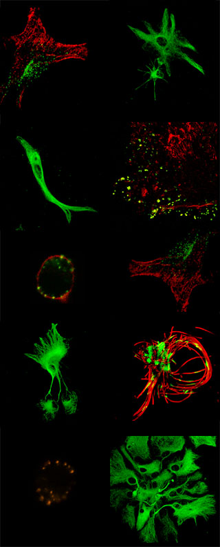
Quantitative analysis of confocal
fluorescence microscopy images
Fluorescent proteins like
green fluorescent proteins (GFP) are markers of
subcellular protein location and trafficking.
After transfection of GFPs, cells or tissue is
imaged. Together with methods of quantitative
image analysis confocal microscopy provide a
powerful tool in molecular cell
biology-physiology studies.
We
developed several software tools, which are now
available
on-line.
References of the
Confocal Center

Sponsor:

|